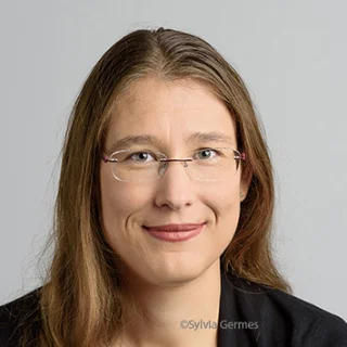
Profile of the supervisor
Rifka Vlijm performed her PhD in the group of Prof.dr. Cees Dekker at the Bionanoscience department of the technical university Delft. Next, she moved to the group of Nobel laureate Prof.dr. Stefan Hell, enabled by both an EMBL long-term fellowship (molecular biology) and a Rubicon grant (proposal ranked 1, physics). Here she built a Stimulated Emission Depletion (STED) microscopy, and developed the first protocols for live-cell STED microscopy on centromeres. In 2019 she joined the Zernike Institute for Advanced Materials as tenure-track assistant professor. In 2022 she received a personal NWO-M1 grant, a personal FSE research grant, and as consortium partner an NWO-XL grant. Furthermore, she was also nominated for the Aspasia price (outcome expected spring 2023). At the national level she is member of the advisory board of the NWO work council “Advanced Methods, Data, and Analyses to understand Living Systems” of the Life Science research community, and of the work council Physics for Life of the Physics community. Since 2021 she is member of the organizational committee of the NWO Dutch Biophysics, of which she was chair for the 2023 conference. She furthermore was involved in the initiation of APNet, the national network for assistant professors.
Expertise
Super resolution microscopy (STED), Live-cell imaging, Cell division
Profile of the research group
In the lab of Rifka Vlijm STED Microscopy is used to study cellular protein structures, especially those involved in cell division. An important part of the research focuses on microscopy developments enabling live-cell imaging: automation for high-throughput measurements and analysis, machine-learning enabled non-toxic imaging, and live-cell imaging protocols. STED microscopy is a powerful technique enabling to reach resolutions of up to 27nm in cells using fluorescence microscopy. Although widely applied to e.g. mammalian cells, standard protocols for live cell yeast and archaea imaging were not yet developed. The Vlijm lab (further) develops labelling and imaging methods to enable STED microscopy in living yeast and archaeal cells, and provides guidelines for sample improvements, to make this technique accessible to a broader community.
